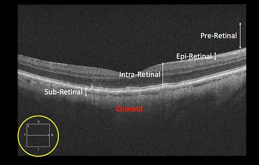
For ease of interpretation, the vitreoretinal anatomy is divided into five unique zones:1. Pre-retinal zone.2. Epi-retinal zone.3. Intra-retinal zone.4. Sub-retinal zone.5. Choroid zone.