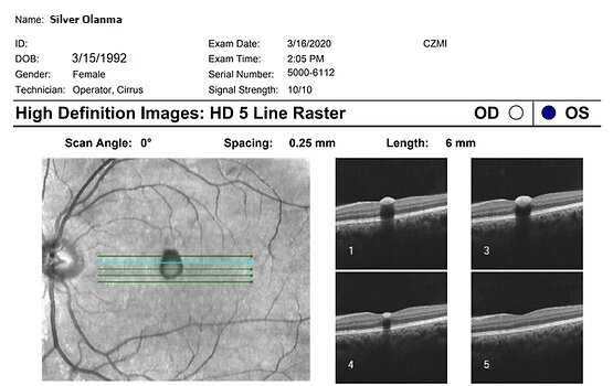
This picture is an OCT sample result. For instance, the Signal Strength of this result is 10/10. Can you see the patientu2019s name, date of birth and Gender?u00a0
What other important demographic information do you see? Macular OCT images are accompanied by a legend to indicate its anatomical orientation (ie. nasal-temporal/superior-inferior).
Remember, the macula is temporal (lateral) about the optic disc. This fact helps the clinician to identify whether the OCT scan represents the OD or OS.
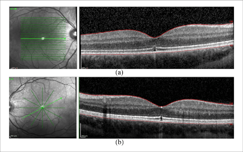
Now, take another look at the OCT scan result below.
Can you classify the image below as OD or OS?
Helpful Tip: The direction of the thicker Retinal Nerve Fibre Layer from the macula hints at which eye you are examining.
The image below is the OD!
Here is the answer: the first image is the OD while the lower image is the OS.
To differentiate between these, note the thicker Nerve Fiber Layer in the upper image.
It is towards the right corner of the image. That is the OD. The Nerve Fiber layer is the hyper-reflective portion at the extreme top of the image.
Notice that in the lower image, the hyper-reflective/whitish layer is towards the left. This is the OS.