When reviewing an OCT scan,u00a0 it is important to describe the structures using the right clinical terminology.
Lighter structures on the OCT image are described as hyper-reflective. Their darker variants are described as hypo-reflective.
For instance,u00a0 structures that assume a Hyper-reflective image result includes blood,u00a0 drusen,u00a0 cotton-wool spots,u00a0 or inflammatory cells.
Some hypo-reflective structures on the OCT scan include fluid,u00a0 vitreous cavity,u00a0 intra-retinal fluid,u00a0 subretinal fluid,u00a0 or blockage of light transmission.
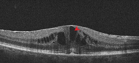
Look at the image below. In this figure, you can see dark irregular structures. The red arrow is pointing at it. This portion of the image is darker than the surrounding retinal tissues. It is described as hypo-reflective.
By the way, the hypo-reflective area is cavities within the retina. It is a fluid-filled pathological condition. If this is happening just below the macular, then macular oedema is confirmed.