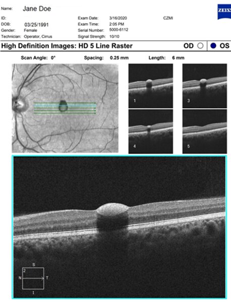
Examining the inner retina, there is a well-delineated, hyper-reflective and concaved area in the inner retina. This lies just posterior to the internal limiting membrane.
This area of hyper-reflectivity is well-defined and could represent haemorrhage, fibrosis or infiltration. In this case, this is a sub-internal limiting membrane haemorrhage.
In the intra-retinal space, there is an area of hypo-reflectivity immediately underneath the hyperreflective lesion on the OCT.
This represents a blocking effect or a shadowing effect from the overlying hyper-reflective material. In the para-foveal area, the layers of the retina can be well visualized and are normal.
There are no obvious abnormalities in the sub-retinal and sub-RPE space. The choroid also appears normal in contour and size.